MCQ : Nuclear-19
A 67 yo man with previous MI and revascularized with CABG. EF 45%.
After new onset of effort angina, he was submitted to dynamic stress rest MPS.
Which is the best interpretation of these images? Look at the s/r MBF and MFR.
A) Normal s/r MBF and normal MFR
B) Regional s/r MBF abnormalities, normal MFR
C) Normal s/r MBF, abnormal MFR
D) Abnormal s/r MBF, abnormal MFR


Correct Answer is D) : Abnormal s/r MBF, abnormal MFR
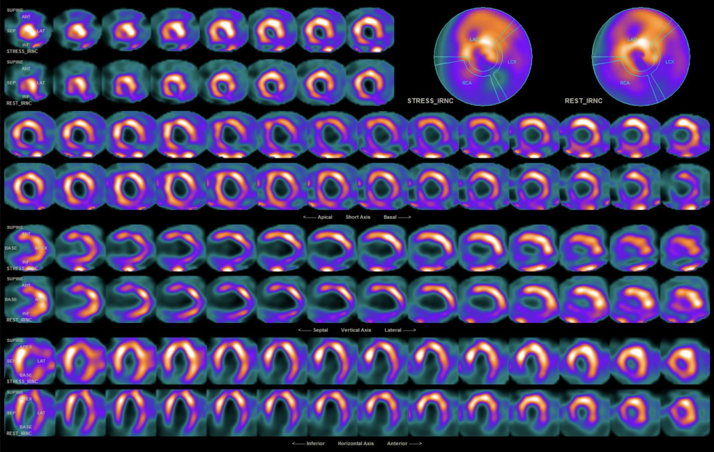
In these pts the MPS scans demonstrated the presence of previous inferior and infero-apical MI, with mild ischemia in the infero-septal wall. The images showed the presence of small necrotic area in the antero-septal wall, middle portion.
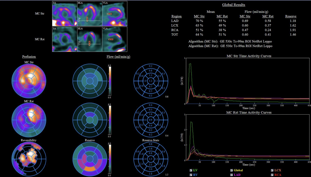
The evaluation of absolute MBF indicated the presence of reduced regional MBF in the three main territories, with regional and global reduced MFR. This information is very important, because the reduction of both MBF and MFR increases the risk of MACE.
![]()
